How ElutiGLASS works
ElutiGLASS is a thin film with two layers. It’s made of PLGA, a material that slowly breaks down in water. The same kind of material is used in absorbable stitches. Each layer is designed to carry one of the two drugs. Here’s how it’s made, how the drugs came out in lab tests, and where it was placed in rabbits.
An electron microscope zooming from 25 to 12,000 times on glass beads 10 µm across. This footage isn’t of ElutiGLASS.
 Video: Zephyris, via Wikimedia Commons
Made in humid air
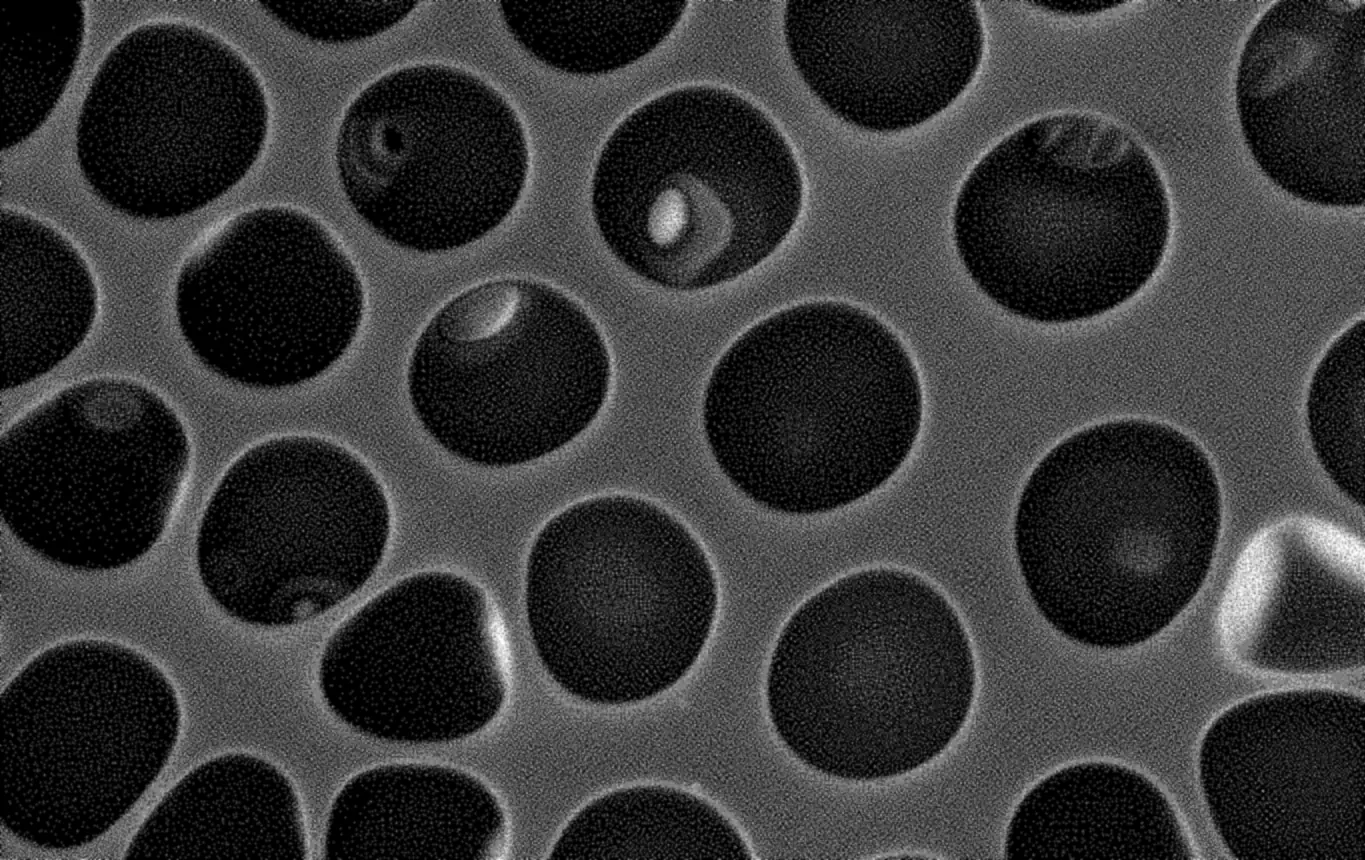
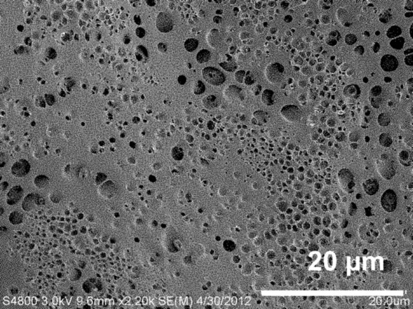
The film is cast in humid air. Tiny water droplets settle on the drying polymer and leave a field of pores, each a few micrometers across. The pattern is called a breath figure. Those pores are where the mitomycin C goes.
Surface pores of an exemplary breath-figure PLGA film. US Patent 10,058,640, Fig. 3.
Two layers, two drugs
First, a lower layer is spun from PLGA mixed with 5-fluorouracil (5-FU), in humid air. A thin sealing layer of a second PLGA goes over it. Mitomycin C is then loaded into that layer’s surface pores. The finished films are sterilized with ultraviolet light.
Schoenberg et al., Translational Vision Science & Technology, 2015, Methods; Swann et al., Journal of Glaucoma, 2019; US 10,058,640, claims 1 and 9.
How long each part lasts
In the lab, test films sat in fluid and were sampled over the following days to see how fast each drug came out. In rabbits, no film was left at three months. Each bar uses the same 90-day scale.
Mitomycin C first day, lab
5-fluorouracil day 3 to 25, lab
The film gone by three months, rabbits
A lab timeline: the dates count the lab and rabbit timings from the chosen day. ElutiGLASS hasn’t been studied in people, and these dates aren’t a prediction for any patient. Sources: US 10,058,640, Figs. 27 and 32; Schoenberg et al. 2015; Swann et al. 2019.
Release, day by day
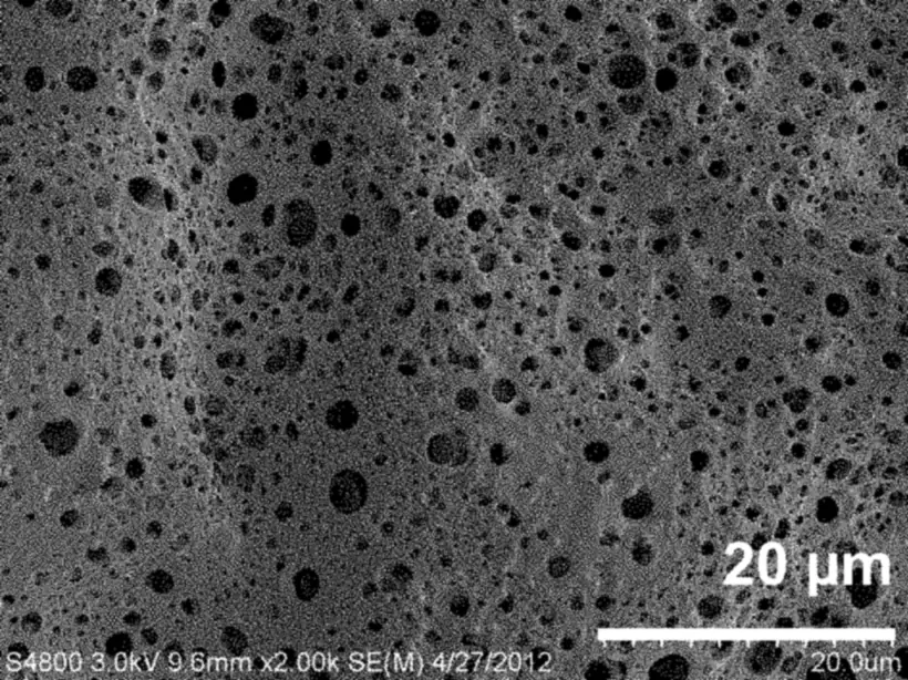
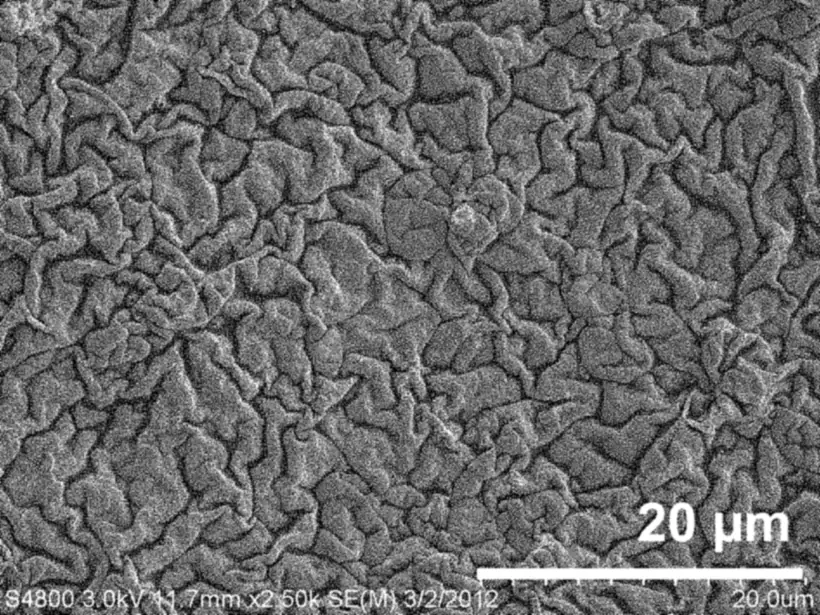
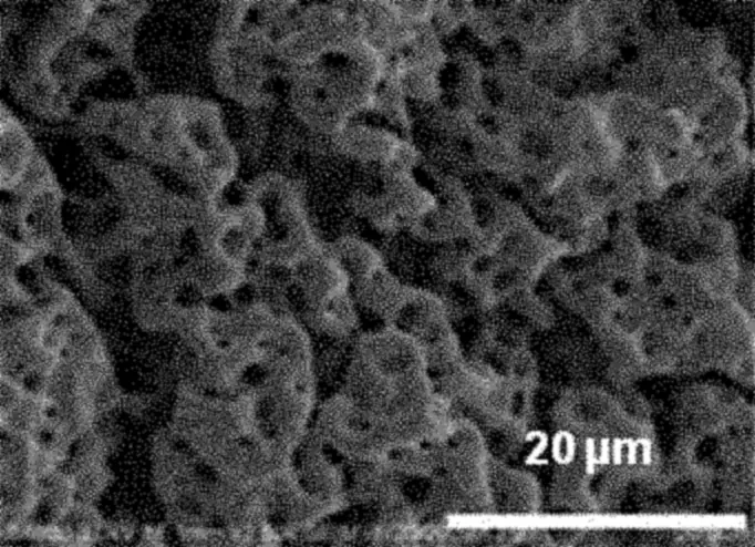
Pick a day to see the film’s surface under an electron microscope and how much of each drug had come out. Everything here was measured in the lab on test films (US Patent 10,058,640). Their doses differ from the real film’s, so each reading names its film.
No image. Both rabbit studies report no film left at three months.
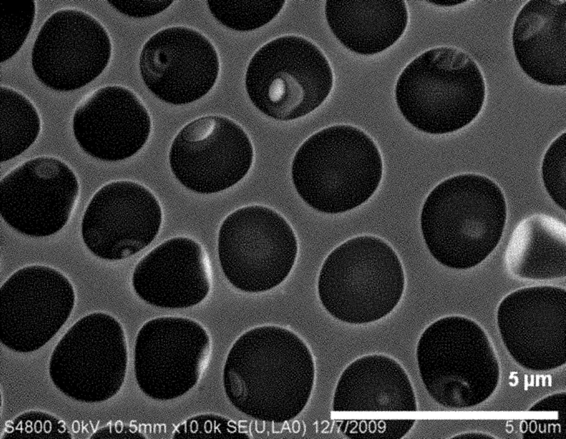
Surface pores of an exemplary breath-figure PLGA film. US Patent 10,058,640, Fig. 3.
Day 0
Day 0. The test films go into fluid.
Mitomycin C, 5 µg film0%
5-FU, 2 mg film0%
- Mitomycin C, 5 µg film about a third by day 1, then level
- Mitomycin C, 1 µg film under a tenth by day 1, about a quarter by day 8
- 5-FU, 2 mg film rises to about three quarters, leveling off near day 25
The values read from the curves
| Day | Released |
|---|---|
| 0 | 0% |
| 1 | 33% |
| 2 | 33% |
| 3 | 35% |
| 4 | 35% |
| 5 | 33% |
| 6 | 32% |
| 7 | 32% |
| 8 | 31% |
| Day | Released |
|---|---|
| 0 | 0% |
| 1 | 8% |
| 2 | 10% |
| 3 | 16% |
| 4 | 21% |
| 5 | 21% |
| 6 | 20% |
| 7 | 22% |
| 8 | 24% |
| Day | Released |
|---|---|
| 0 | 0% |
| 1 | 2% |
| 2 | 4% |
| 3 | 6% |
| 4.5 | 10% |
| 6 | 17% |
| 7 | 28% |
| 8 | 38% |
| 10 | 45% |
| 13 | 48% |
| 14 | 54% |
| 16 | 59% |
| 22 | 74% |
| 25 | 76% |
| 30 | 76% |
| 33 | 76% |
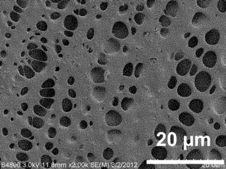
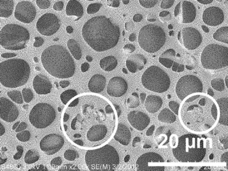
Mitomycin C: share released from 5 µg and 1 µg test films (Fig. 27), measured to day 8. 5-FU: share released from a 2 mg test film with no mitomycin C (Fig. 32), measured to day 33. Values are read from the published curves and are approximate. A whole ElutiGLASS film carries about 1.3 µg of mitomycin C and 0.9 mg of 5-FU (Swann et al., 2019, Methods); its own release hasn’t been published. Images: an exemplary breath-figure PLGA film (Fig. 3), samples of one two-layer test film type carrying 0.4 mg of 5-FU (Fig. 35), and a breath-figure PLGA test film at day 35 (Fig. 10).
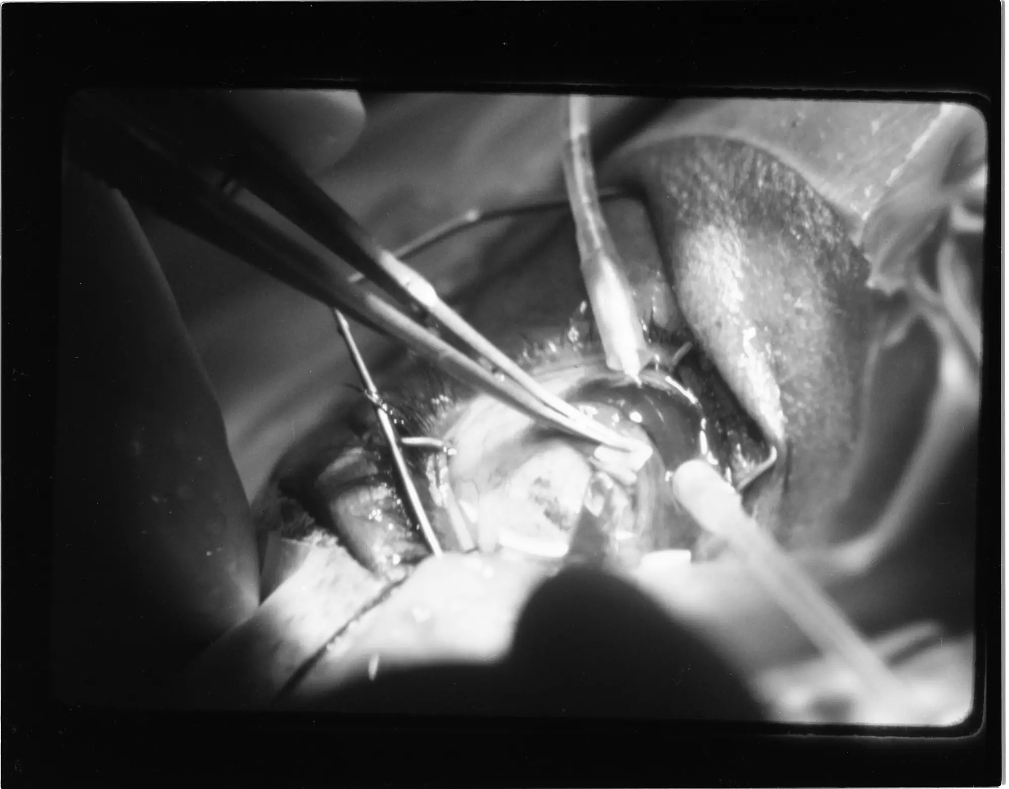
Where the film is placed
Glaucoma surgery at the Kresge Eye Institute, Detroit, 1978. NASA Glenn Research Center, via the U.S. National Archives, public domain.
Trabeculectomy
The surgeon cuts a small trapdoor in the white of the eye, called the scleral flap. Fluid drains out under it. The film was placed under the conjunctiva, the thin membrane over the white of the eye. About a quarter of it lay over the flap, and the rest behind it. Studied in rabbits (Swann et al., 2019); see the results.
Glaucoma drainage devices
A small tube carries fluid out of the eye to a plate on its outer wall. The film is designed to sit on that plate, as on the Ahmed valve. Studied on Ahmed valves in rabbits (Schoenberg et al., 2015); see the results.
Shunts and microshunts
The company also lists the ExPRESS shunt and microshunts such as the InnFocus MicroShunt, now PreserFlo. These uses haven’t been studied.