Contact

Evidence

What’s known about ElutiGLASS so far comes from two studies in rabbits and from lab tests. No study in people has been published yet. Each result names its study, and the gaps are listed after the results.

  1. Rabbit studies
  2. Lab tests
  3. Compared with current practice
  4. What hasn’t been studied
  5. Papers and patents

Results in rabbits and the lab

19

rabbits, trabeculectomy, three months

Swann et al., Journal of Glaucoma, 2019

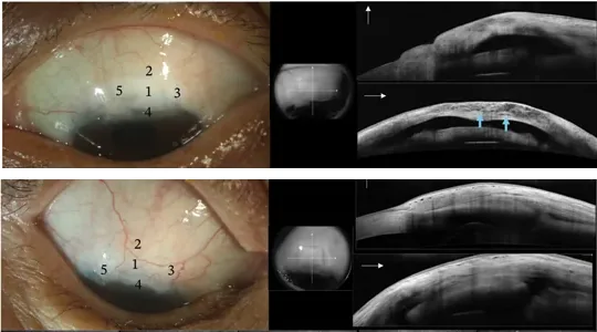
Two human eyes after trabeculectomy with cataract surgery, each shown in a slit-lamp photograph and two OCT scans: above, a functioning filtering bleb; below, a non-functioning one.
Filtering blebs in people, for comparisonAbove, a functioning filtering bleb; below, a non-functioning one, each in a slit-lamp photograph and OCT scans. From a study of trabeculectomy with cataract surgery; ElutiGLASS wasn’t used. Lai et al., Journal of Ophthalmology, 2026, Fig. 1, cropped. CC BY 4.0

Two film sizes, compared with mitomycin C on a sponge

In a trabeculectomy, fluid drains into a small pocket under the eye’s surface, called the bleb. The question is whether it stays open. The film was cut to a quarter or a half of its full size, and compared with the usual mitomycin C (MMC) on a sponge. The authors note that the eye-pressure differences between groups were near the range of error for their handheld pressure gauge (Tono-Pen XL) in these rabbits.

A quarter of the film.
5-FU
0.23 mg
Mitomycin C
0.33 µg
Fibrosis, median
1

Slow release, designed for 23 to 30 days. 5 of 6 eyes kept a bleb space, with lower, more diffuse blebs. Neither bleb space nor fibrosis differed significantly from the sponge.

Eyes with a bleb space at three months

Sponge, MMC 0.4 mg/mL4 of 6
Quarter film5 of 6
Half film0 of 7

Sponge group: 2 of 6 rabbits died at one month, for unknown reasons; all 4 examined at three months kept a bleb space. The quarter film’s 5 of 6 wasn’t significantly different from the sponge. Fibrosis, the scarring, was scored from 1 to 5 by looking at the tissue under a microscope (histology).
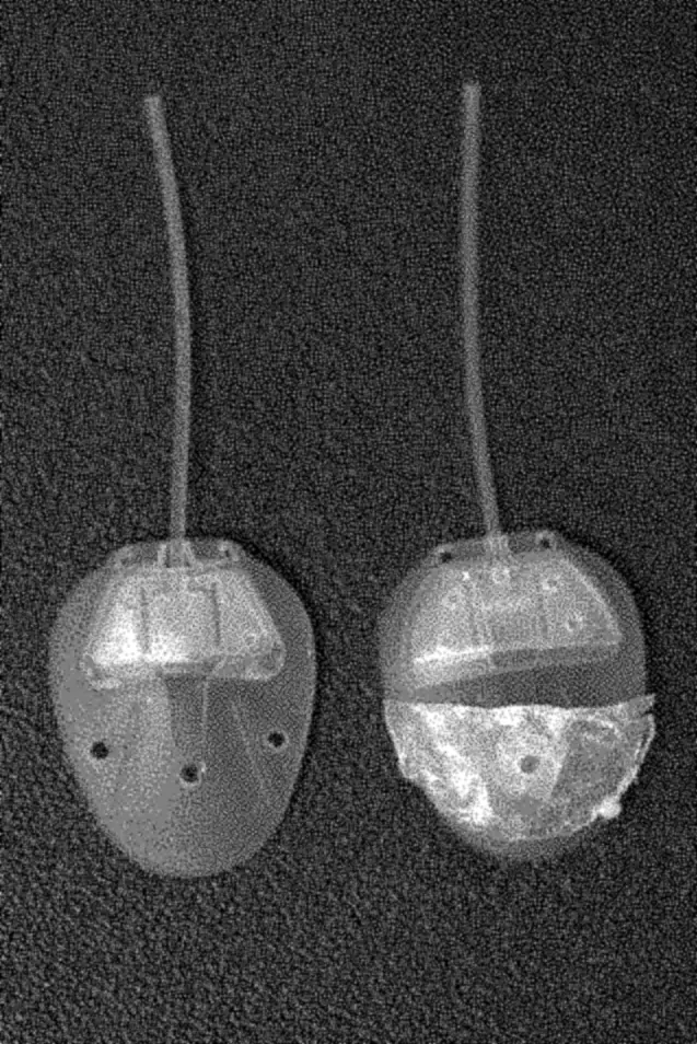
Photograph of two Ahmed glaucoma valves: a commercial valve, and one coated with a breath-figure PLGA film.
A film-coated Ahmed valveLeft, an Ahmed valve as sold; right, one coated with breath-figure PLGA film. US 10,058,640, Fig. 20.

On drainage implants: thinner tissue over the drainage pocket

The films sat on Ahmed valves, a kind of drainage implant. A bleb forms over the implant’s plate. Over films with 5-fluorouracil (5-FU) and mitomycin C, the tissue roof over that pocket measured 316 to 320 µm. Over a drug-free film, it measured 544 µm. That difference is very unlikely to be chance (P < 0.001). Scarring scores under the microscope didn’t differ between groups (P = 0.14).

Bleb roof thickness, mean, µm

PLGA film, no drug544
Hydrogel disk, 6.5 µg MMC373
PLGA, 0.45 mg 5-FU + MMC316
PLGA, 1.35 mg 5-FU + MMC320

Each film with both drugs carried 0.65 µg of mitomycin C. Not shown: films with 5-FU alone (0.45 mg and 1.35 mg), which the authors found not effective in reducing fibrosis.

Lab

test films, in fluid and with cultured cells

US Patent 10,058,640, Figs. 25 to 28, 32 and 36

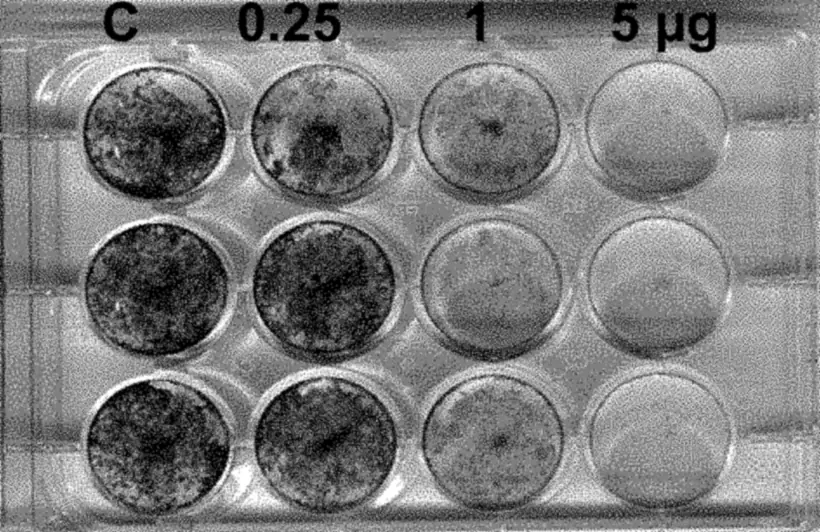
A 12-well culture plate after staining: wells under a plain film, marked C, stay dark with cells; wells under films with 0.25, 1 and 5 µg of mitomycin C are paler, the 5 µg wells palest.
Stained cells: darker wells hold more of them. C is a plain film. US 10,058,640, Fig. 25.

In the lab: release, cell tests and breakdown

A 5 µg test film released about a third of its mitomycin C on the first day and little more by day 8. A 2 mg test film released its 5-FU after a delay of a few days from the sealing layer, leveling off near day 25 at about three quarters. The curves, day by day.

In a cell test, films carrying 0.25, 1 and 5 µg of mitomycin C killed about 10%, about 60% and nearly 90% of the cells, compared with a plain film. The cells were COS-1, a standard lab cell line. What a 5 µg film released on the first day killed about 88% of them. What it released between days 4 and 8 killed about 8%.

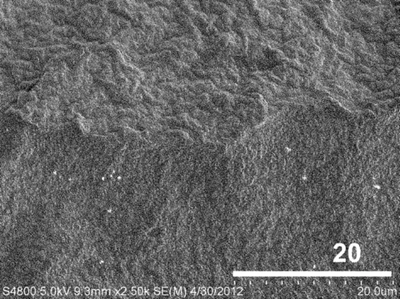
Cross-sections of a two-layer test film show the top layer’s pores at day 7, and none by day 28, when the surface had wrinkled.

Electron micrograph of a two-layer test film's cross-section after 28 days in fluid: a wrinkled surface with no open pores.
A two-layer test film after 28 days in fluid, in cross-section. US 10,058,640, Fig. 36.

Compared with current practice

Surveyed American Glaucoma Society members used mitomycin C in 97% of primary trabeculectomies in 2016, most often on a sponge; 31% injected it instead.

Vinod et al., Journal of Glaucoma, 2017 (the 2016 survey).

Measure Sponge-applied mitomycin CCurrent practice ElutiGLASSInvestigational
Drugs Mitomycin CMitomycin C, then 5-fluorouracil
Dose Varies with the sponge and the surgeon. Hand-cut sponges soaked at 0.2 mg/mL held 1.9 to 17.3 µg. Mehel et al., Journal of Ocular Pharmacology and Therapeutics, 1998 Set by the piece of film used: about 1.3 µg of mitomycin C and 0.9 mg of 5-FU in a whole film. Swann et al. 2019, Methods
Duration Applied for 45 seconds to 4.5 minutes, then removed. AGS survey, 2016 Designed to release mitomycin C in the first day, and 5-FU from day 3 to 5 for 23 to 30 days. Swann et al. 2019; US 10,058,640, claims 2 to 4
What remains No material is left; the sponge is removed. The film stays and breaks down. None was found at three months in rabbits. Schoenberg et al. 2015; Swann et al. 2019
Evidence in people Decades of clinical use, with randomized trials and Cochrane reviews. None yet. Rabbit and laboratory studies only.

What hasn’t been studied

What these studies can’t tell you yet.

  • Use in peopleNo study has been published, and ClinicalTrials.gov lists none (searched Oct 19, 2026).
  • Release inside a living eyeEvery release figure on this site comes from lab tests, with films sitting in fluid. No published study has measured how fast the drugs come out of the ElutiGLASS film inside an eye, in rabbits or in people.
  • The ElutiGLASS film’s own releaseThe published release curves come from test films with other doses (US 10,058,640, Figs. 27, 31 and 32). The release of the film itself hasn’t been published.
  • Shunts and microshuntsThe ExPRESS shunt and the microshunts the company lists haven’t been tested with the film.
  • Beyond three monthsBoth rabbit studies ended at three months, so there’s no animal data beyond that.

That part of the page isn’t here

The link pointed to a section that isn’t here. The closest matches: