
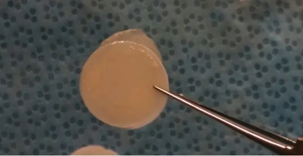
ElutiGLASSA dissolving film for glaucoma surgery
Designed to release two anti-scarring drugs in turn, from one piece placed during surgery. Investigational and not approved by the FDA.
See how it worksPlaced during surgeryIn the 2019 rabbit study it went under the conjunctiva, the eye’s thin outer membrane, with about a quarter of it over the scleral flap.
Three months later, neither rabbit study found any of the film left.
Why glaucoma surgery can stop working
Glaucoma surgery lowers eye pressure by giving fluid a new way out of the eye. Two common kinds are a trabeculectomy and a drainage implant. But the eye heals. Scar tissue can close that new outlet, and then the pressure rises again. To slow that healing, surgeons apply mitomycin C (MMC) during the operation. Some also inject 5-fluorouracil (5-FU) in the weeks after. Both drugs slow the cells that form scar tissue.
Wilkins et al., 2005, and Green et al., 2014, Cochrane Database of Systematic Reviews; Budenz et al., Ophthalmology, 2015.
ElutiGLASS is designed to deliver both drugs from one film at the outlet, over about four weeks. Whether it changes outcomes in people hasn’t been studied.
How often the surgery had failed after five years, in four randomized trials
Ahmed and Baerveldt are two kinds of drainage implant. Failure means pressure too high or too low, another glaucoma operation, or loss of light perception. AVB used a stricter pressure target. TVT, PTVT, ABC and AVB five-year reports (PMIDs 22245458, 35835337, 25439606, 27544023).
Two drugs, one after the other
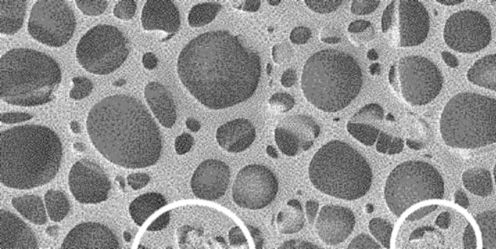
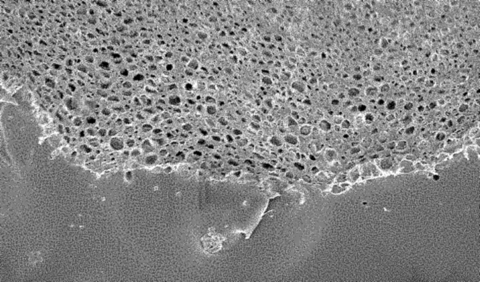
The film has two thin layers. The top one carries mitomycin C in its tiny surface pores. It’s designed to come out within the first day. The layer beneath holds 5-fluorouracil. It’s designed to start a few days later (day 3 to 5) and to last three to four weeks (23 to 30 days).
Amounts in a whole 13 mm film and timing as reported by Swann et al., Journal of Glaucoma, 2019, Methods. Model drawn from US 10,058,640, claim 1 and Figs. 29 and 36, and colored after ElutiMed’s photograph of the film. Thickness and pores enlarged.
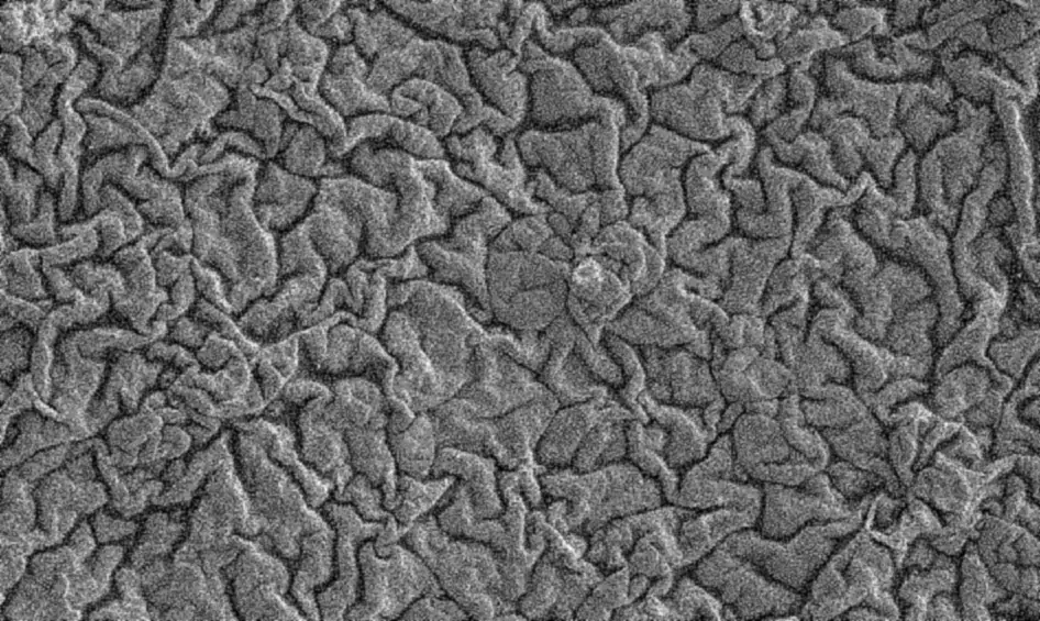
Micrographs: US 10,058,640, Figs. 29, 35 and 36. Photograph: ElutiMed.
How the film is cast and tested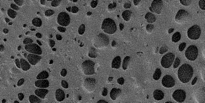
The film in the eye
In a trabeculectomy, the surgeon cuts a small trapdoor in the white of the eye. It’s called the scleral flap. Fluid drains out under it and collects in a small blister called the bleb. The bleb sits under the conjunctiva, the eye’s thin outer membrane. In the 2019 rabbit study, the film went into that space. About a quarter of it lay over the flap, and the rest behind it.
Day 0, at surgery
- Day 0 In the rabbit study, a quarter or half of a 13 mm film was placed at surgery
- Day 1 Mitomycin C’s first-day release, in a lab test
- Day 255-FU release levels off, in a lab test
- 3 months No film left, in both rabbit studies
Schematic and not to scale. Placement as described in Swann et al., Journal of Glaucoma, 2019. The timing comes from lab tests (US 10,058,640, Figs. 10, 27 and 32) and the design goal of breaking down in 2 to 3 months. No published study has measured release from the ElutiGLASS film inside an eye. Both rabbit studies (Schoenberg et al. 2015, Swann et al. 2019) report the film gone at three months.
What has been studied
Two studies in rabbits, and lab tests of how the film releases its drugs and breaks down.
-
Trabeculectomy19 rabbits, three months
Rabbits got a quarter or a half of a film, or the usual mitomycin C on a sponge. Five of six eyes with a quarter film kept their bleb, the drainage pocket. None of the seven with a half film did.
Swann et al., Journal of Glaucoma, 2019
-
Ahmed valvesA drainage implant; 48 rabbits, three months
With films carrying both drugs, the tissue over the drainage pocket was thinner than with a drug-free film. Scarring scores under the microscope didn’t differ significantly.
Schoenberg et al., Translational Vision Science & Technology, 2015
-
PeopleNot yet studied
Nothing published, and no trial registered on ClinicalTrials.gov.
Searched Oct 19, 2026
ElutiGLASS in 90 seconds
What the film says, with its sources
- ElutiGLASS. A dissolving film for glaucoma surgery.
- Glaucoma surgery lowers eye pressure by giving fluid a new way out of the eye. But the eye heals, and scar tissue can close that opening, so the pressure rises again. (Wilkins et al., 2005; Green et al., 2014. Cochrane Database of Systematic Reviews.)
- To slow that healing, surgeons use mitomycin C during the operation, and sometimes 5-fluorouracil afterwards. (Same sources.)
- ElutiGLASS is a dissolving film designed to deliver both drugs from one piece, placed during surgery. Investigational. Not approved by the FDA.
- Cast in humid air, its surface is covered in tiny pores that hold the mitomycin C. (An exemplary breath-figure PLGA film, US 10,058,640, Fig. 3; mitomycin C loaded into the pores, claim 1.)
- The 5-fluorouracil sits in a second layer underneath, designed to come out over the following weeks. Top layer: mitomycin C, about 1.3 µg in a whole film. Lower layer: 5-fluorouracil, 0.9 mg in a whole film. (US 10,058,640, Fig. 29; amounts and timing as reported by Swann et al., Journal of Glaucoma, 2019, Methods.)
- In lab tests, mitomycin C came out on the first day, and 5-FU started to follow a few days later. A 5 µg test film released about a third of its dose on day 1; a 2 mg test film released about three quarters, leveling off near day 25. (Test films with other doses; US 10,058,640, Figs. 27 and 32.)
- In fluid, its surface changes week by week, until the pores close: days 7, 14 and 28. (Samples of one two-layer test film type, 0.4 mg 5-FU; US 10,058,640, Fig. 35.)
- In rabbit studies, the film was placed during surgery, under the thin membrane over the white of the eye: under the conjunctiva, about a quarter over the scleral flap. (Swann et al., Journal of Glaucoma, 2019. Schematic, not to scale.)
- Three months later, neither rabbit study found any of the film left. (Swann et al., 2019; Schoenberg et al., Translational Vision Science & Technology, 2015.)
- ElutiGLASS is investigational. It hasn’t been studied in people, and it isn’t approved by the FDA. ElutiMed, Inc., Tampa, Florida.
Team

Ramesh S. Ayyala, MD
Founder
Professor and Chair of Ophthalmology at the USF Morsani College of Medicine, and a co-inventor of ElutiGLASS.

Diana Shapiro
Chief Business Officer
More than 23 years building companies, about a decade of it turning university research into businesses.
Contact ElutiMed
Questions about the research, a partnership or an investment.
Write to ElutiMed